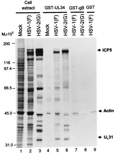
FIG. 6.
Autoradiographic images of electrophoretically separated infected cell proteins bound to GST or to GST-UL34 fusion protein. HEp-2 cells were infected and labeled with radioactive methionine as described in the legend to Fig. 2. The protein complexes bound to beads were subjected to electrophoresis on an SDS–8% polyacrylamide gel, transferred to a nitrocellulose membrane, and subjected to autoradiography. Lanes 1 to 3, lysates of mock-infected cells or cells infected with HSV-1(F) or HSV-2(G), respectively; lanes 4 to 6, proteins pulled down by GST-UL34 chimeric protein from cells mock infected or infected with HSV-1(F) or HSV-2(G), respectively; lanes 7 to 9, proteins pulled down by GST-gB fusion protein (lanes 7 and 8) or just GST (lane 9). Arrows indicate the infected cell proteins specifically precipitated by GST-UL34. The identities of UL31 and ICP5 were verified by immunoblotting.

