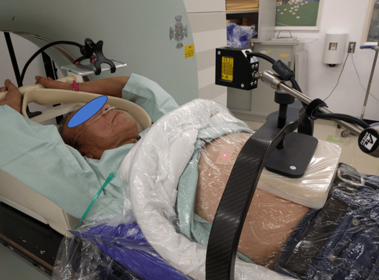
Figure 1. A photograph showing a CT simulation for lung tumor treatment planning under abdominal compression using a vacuum-assisted fixation device and an in-house compression block with a carbon fiber frame to apply pressure to the block.
A narrow laser beam was projected perpendicular to the patient's abdomen with an aimed laser-abdomen distance of 12 cm. The laser spot was projected on the subcostal line midway between the inferiormost thoracic cage border and the L3 vertebral body to detect respiratory motion while minimizing the effect of the aortic pulsation. The reflected light was detected on a 1D array sensor, where the detected signal position indicates the distance to the abdomen. The laser sensor detects abdominal displacement caused by breathing and generates a respiratory curve, which was then exported to the CT unit to reconstruct multi-phase 4D CT images, where amplitude-based respiratory phase information was obtained from the respiratory curve. A portable display was placed over the patient's face to provide visual guidance for stable breathing. Treatment plans were created using the end-expiratory CT images.
CT: computed tomography

