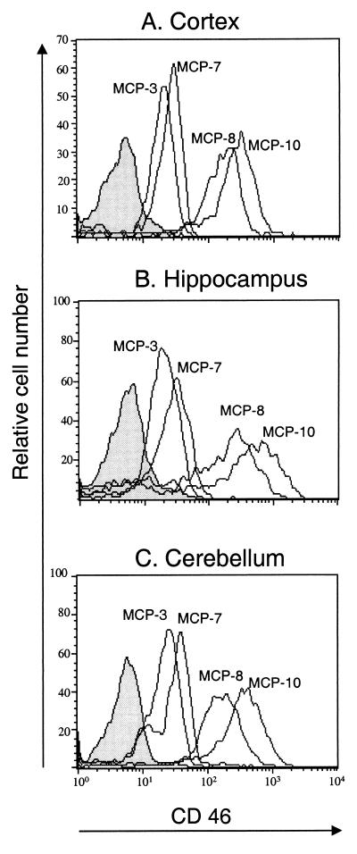
FIG. 1.
CD46 expression in the brains of transgenic mice. Brain structures were dissected from PBS-perfused CD46 C-Cyt1 transgenic (MCP-8 and MCP-10), CD46 C-Cyt2 transgenic (MCP-3 and MCP-7), and nontransgenic (BALB/c) mice. Frontal cortex (A), hippocampus (B), and cerebellum (C) were gently dissociated and stained with anti-CD46 rabbit polyclonal serum, followed by FITC-conjugated goat anti-rabbit IgG. Dead cells were excluded by propidium iodide staining. Negative control histograms (nontransgenic mice) are filled in gray. Results are representative of three different experiments.

