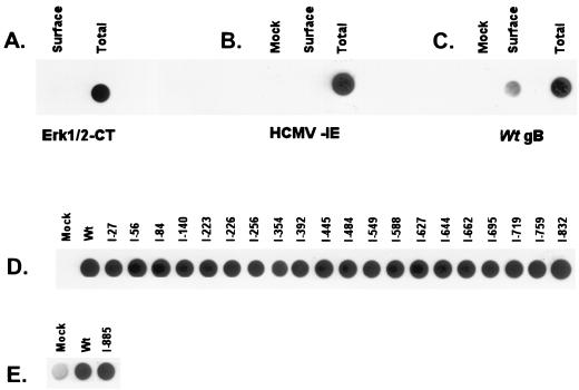
FIG. 5.
Cell surface detection of the gB insertion mutants by using a biotinylation assay. Transfected cells were biotinylated, then lysed in a 1% CHAPS solution, and precipitated with streptavidin-agarose. The biotinylated proteins were boiled in 2× sample buffer and transferred to nitrocellulose. The biotinylated proteins were then detected with antibodies described in Materials and Methods. Total amount of protein in the cells was detected essentially the same, except that the cells were lysed prior to biotinylation. (A) Control examining both cell surface and total expression of the cytoplasmic protein Erk1/2; (B) control examining both cell surface and total expression of the transfected HCMV IE protein; (C) control using wt gB examining both cell surface and total expression (MAb 58-15 was used for detection; data were quantitated with a Bio-Rad phosphorimager); (D) cell surface expression of the insertion mutants, using MAb 58-15 for detection; (E) cell surface expression of I-885; MAb 3C-2 was used for detection because MAb 58-15 reacts poorly with I-885 (Fig. 2).

