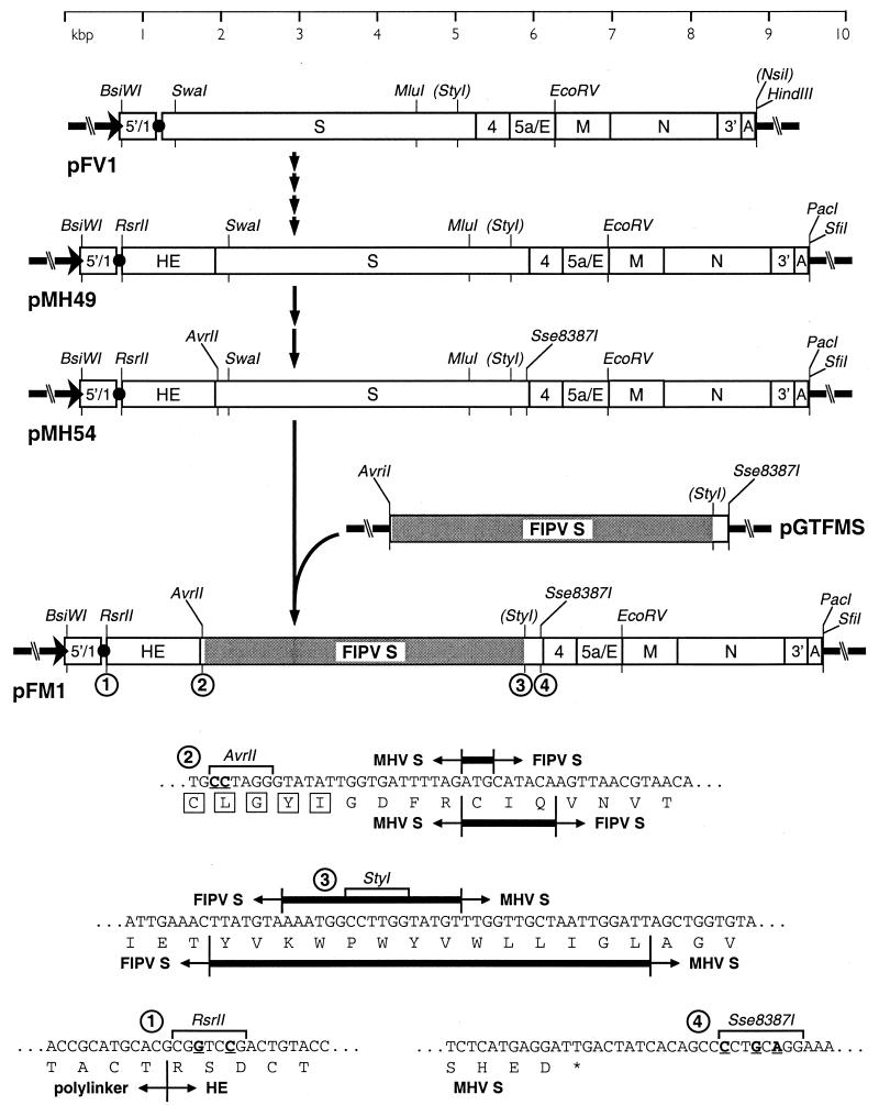
FIG. 1.
Construction and composition of the donor RNA template for incorporation of the FIPV S gene ectodomain into MHV. Transcription vector pFM1 was derived from parent plasmid pFV1 (13) via six intermediates, including pMH49 and pMH54, as described in Materials and Methods. The chimeric FIPV-MHV S gene was shuttled into pFM1 from the subclone pGTFMS. MHV and FIPV sequences are indicated, respectively, by open and shaded rectangles. The arrow at the left end of each vector indicates the T7 promoter; the solid circle represents the polylinker between the 5′-end segment of the MHV genome (denoted 5′/1) and the 3′ region containing the structural genes, the 3′ untranslated region (denoted 3′), and the polyadenylated segment (denoted A). Restriction sites relevant to plasmid construction are shown and, unless enclosed in parentheses, are unique in the plasmid in which they appear. At the bottom are shown the sequences in pFM1: 1, between the polylinker and the HE gene fragment; 2, at the MHV-FIPV junction in the signal peptide-encoding portion of the chimeric S gene (with signal peptide residues boxed); 3, at the FIPV-MHV junction in the transmembrane domain-encoding portion of the chimeric S gene; and 4, in the region immediately downstream of the S gene. Nucleotides mutated to create restriction sites are underlined. The boundaries between MHV and FIPV sequence are indicated by short vertical lines; thicker horizontal bars between these indicate nucleotides or amino acids common to both the MHV and FIPV sequences.

