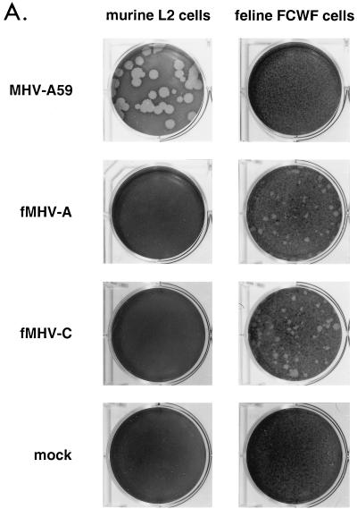
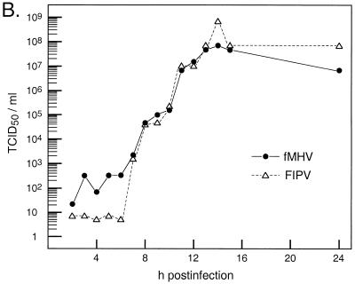
FIG. 3.
Growth of fMHV in feline cells. (A) Plaque-forming ability of fMHV. Monolayers of murine L2 cells or feline FCWF cells were mock infected or infected with wild-type MHV or either of two independent isolates of fMHV. Plaques were visualized at 66 h postinfection, after staining with neutral red. (B) Single-step growth kinetics of fMHV-C and FIPV in FCWF cells. Viral infectivity in culture medium at different times postinfection was determined by a quantal assay on FCWF cells, and 50% tissue culture infective doses (TCID50) were calculated.


