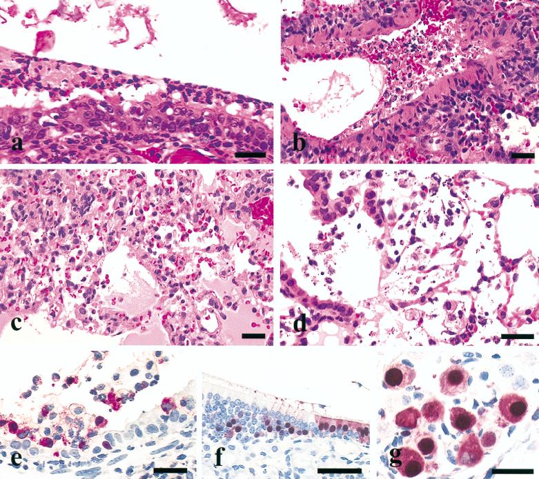
FIG. 2.
Experimental studies in BALB/c mice inoculated intranasally with different H5 avian influenza viruses. Photomicrographs of hematoxylin-and-eosin-stained tissue sections (a to d) or sections stained immunohistochemically to demonstrate avian influenza virus NP (e to g). (a) Disorganization, degeneration, and necrosis of respiratory epithelium of the nasal turbinate with lumenal fibrinopurulent exudate from a mouse euthanatized 4 days after inoculation with HK156 (bar = 25 μm). (b) Necrotizing bronchitis with lumenal hemorrhage and necrotic debris from a mouse euthanatized 4 days after inoculation with HK220 (bar = 25 μm). (c) Peribronchial alveolitis characterized by intra-alveolar serofibrinous exudate, erythrocytes and neutrophils, increased alveolar macrophages, and inflammatory cells in alveolar walls from a mouse euthanatized 4 days after inoculation with HK156 (bar = 25 μm). (d) Necrotizing alveolitis with intra-alveolar macrophages, neutrophils, and fibrin from a mouse euthanatized 4 days after inoculation with HK728 (bar = 25 μm). (e) Avian influenza virus NP in respiratory epithelium, macrophages, and neutrophils in the bronchus from a mouse euthanatized 4 days after inoculation with Eng/91 (bar = 25 μm). (f) Avian influenza virus NP in respiratory (right) and olfactory (left) epithelia of the nasal turbinate from a mouse euthanatized 4 days after inoculation with HK156 (bar = 50 μm). (g) Intranuclear and intracytoplasmic avian influenza virus NP in ganglial neurons adjacent to the bronchial lymph node from a mouse euthanatized 4 days after inoculation with HK156 (bar = 25 μm).

