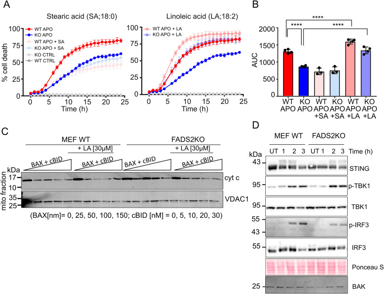
Fig. 5. Membrane unsaturation promotes MOMP and apoptotic cell death.
A Kinetics of cell death (measured as %DRAQ7 positive cells) in WT and FADS2 KO U2OS cells. Cells were fed with lipids (stearic acid, linoleic acid or vehicle) for 18 h, and then treated or not with ABT-737 (10 µM) and S63845 (10 µM) for apoptosis induction. Data are presented as means ± S.D..; n = 4 independent experiments. B Comparison of the samples in (A) by quantification of the area under the curve (AUC). Data are means ± S.D; n = 4; ****, p = 0,0001 by one-way ANOVA corrected for multiple comparisons using Tukey’s multiple comparison test) (C) WB analysis of cytochrome c release from mitochondria isolated from WT or FADS2 KO MEFs fed with lipids or not for 18 h. Isolated mitochondria were treated with increasing amounts of BAX and cBID as indicated for 1 h prior to separation of mitochondrial pellets and supernatants. Cytochome c level in the mitochondrial fraction were then detected by immunoblot using anti-cytochrome c antibody. Representative Western blot of n = 3 independent experiments. D Activation of the STING pathway downstream of MOMP. WT or FADS2 KO MEFs were treated with ABT-737 (5 µM) and S63845 (5 µM) for the indicated times in the presence of pan-caspase inhibitor. Levels of STING, phosphorylated TBK1, and IRF3 were detected by WB. Actin used a loading control. Representative Western blot of n = 3 independent experiments. Source data are provided as a Source Data file.

