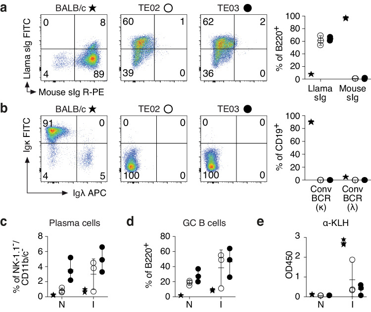
Fig. 2. LamaMice support the development of B cells that display llama heavy chain immunoglobulins.
a, b Spleen cells of 16-27-week-old mice (n = 3 per group) were stained with fluorochrome-conjugated antibodies and analyzed by flow cytometry. Gating was performed on B220+ (a) or CD19+ (b) cells. c–e 12-13-week-old mice (n = 3 per group) were immunized (I) with keyhole limpet hemocyanin (KLH). Naive (N) mice served as controls. c, d Four days after the second boost, spleen cells were stained with fluorochrome-conjugated antibodies and analyzed by flow cytometry. c Gating was performed on NK-1.1/CD11b/CD11c triple-negative cells. Plasma cells were identified as CD138+/PC-1+ cells. d Gating was performed on B220+ cells. Germinal center (GC) B cells were identified as CD95+/surface immunoglobulin (sIg)int cells. e Serum was analyzed for the presence of KLH-specific antibodies (α-KLH) by ELISA using peroxidase-conjugated mouse Ig-specific (BALB/c) or llama Ig-specific (LamaMice) secondary antibodies. a–e Asterisks indicate samples from BALB/c mice, open circles from TE02 LamaMice, closed circles from TE03 LamaMice. a, b Dot plots are from single representative animals. Numbers indicate the % of cells in the respective quadrant. a–e Bar diagrams show the corresponding results for all mice in a group. Data represent mean ± SD for n = 3 individuals.

