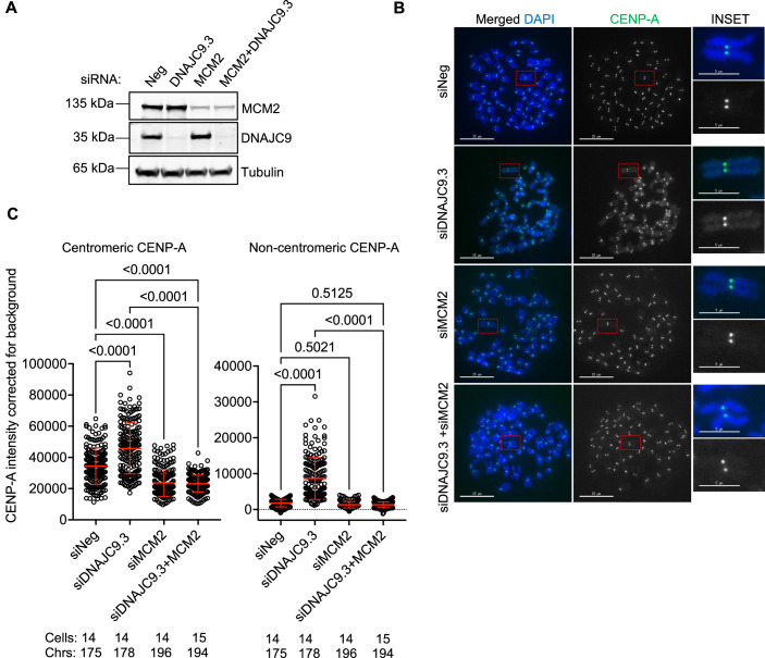
Figure 6. MCM2 contributes to CENP-A mislocalization in DNAJC9-depleted cells.
(A) Western blot of whole-cell extracts prepared from control or DNAJC9-depleted cells with or without MCM2 depletion showing the efficiency of DNAJC9 and MCM2 depletions in HeLa YFP-CENP-ALow. Alpha-tubulin was used as the loading control. Representative images from three biological replicates are shown. (B) MCM2 depletion suppresses CENP-A mislocalization in DNAJC9-depleted HeLa YFP-CENP-ALow cells. Representative images of metaphase chromosome spreads immunostained for CENP-A in control or DNAJC9.3 siRNA-transfected cells with or without MCM2 depletion in HeLa YFP-CENP-ALow from three biological replicates. Scale bar: 15 µm. Scale bar for inset: 5 µm. (C) Scatter plots showing CENP-A intensities corrected for background at the centromeric or noncentromeric regions as described in B. Each dot represents value from the individual chromosome. Chrs represent the total number of chromosomes measured per condition from three biological replicates. Red horizontal lines represent mean signal intensity and error bars represent SD across all measurements from three biological replicates. P values were derived from one-way ANOVA with Tukey’s ad hoc test. Source data are available online for this figure.

