Figure 2.
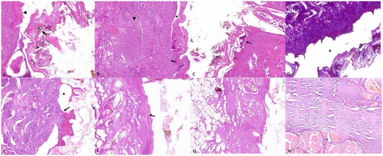
Representative skin pathological alterations in fire-related death cases (Group A and B): (A) soot on the surface of the skin (arrow) and elongation of epithelial cells (arrowhead) (B) inflammatory infiltrate characterized by neutrophilic granulocytes (arrowhead), associated with necrosis (arrow) and epithelial detachment (asterisk) (C) intraepidermal blister (arrow), (D) increase in connective tissue basophilia and dermo-epidermal and intraepidermal separation (asterisk) (E) dermo-epidermal separation (asterisk) and sub-epidermal blister (arrow); Representative skin pathological alterations in experimentally exposed cadavers (Group C): (F) loss of cellular details and dermo-epidermal separation (arrow) (G) dermal vacuolization (H) increased connective tissue basophilia.

