. 2024 Mar 19;84(11):1799–1816. doi: 10.1158/0008-5472.CAN-23-3248

Figure 3.

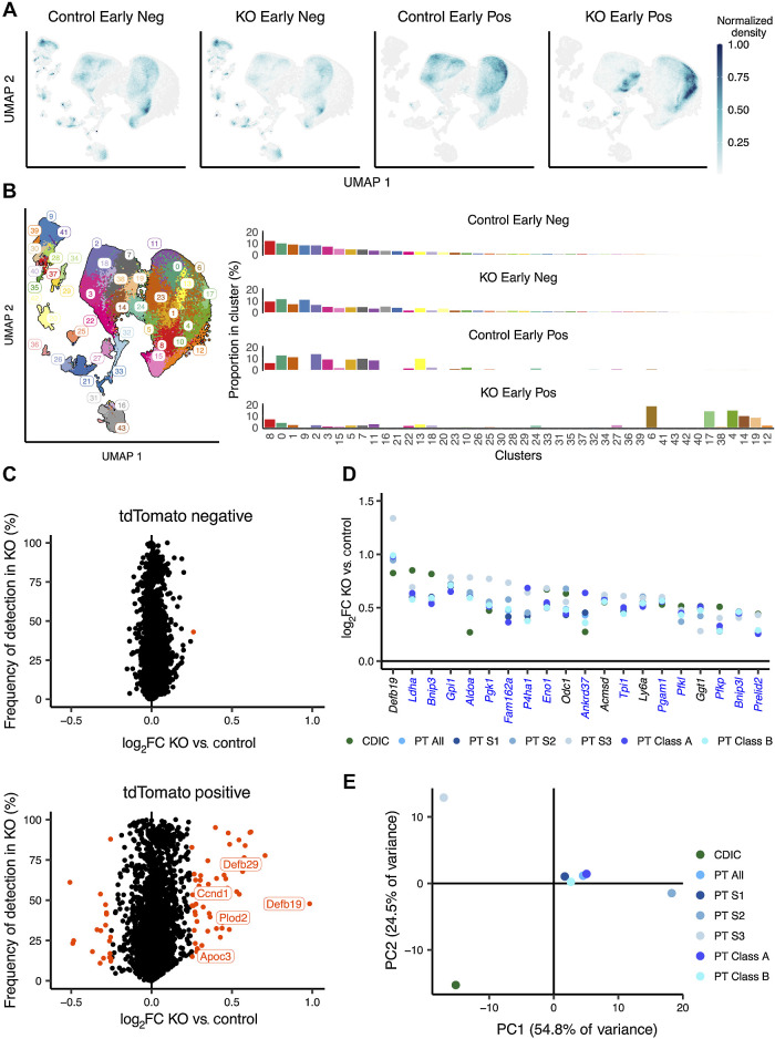
Figure 3. Biallelic Vhl inactivation entrains early cell-specific transcriptomic changes in RTE cells. A, Density plot depicting UMAP distribution of tdTomato-negative and -positive cells from kidneys of Control and KO mice harvested early after recombination. B, Left, UMAP plot depicting cells from Control and KO mice harvested early after recombination colored by UMAP clusters. Right, proportion of cells from each condition belonging to any cluster. C, Scatter plot depicting frequency of expression in tdTomato-negative (top) or tdTomato-positive (bottom) cells from KO mice against log2-fold change (log2FC) between cells from KO versus Control mice for all genes at the early time point. Orange, significantly regulated genes. Genes explicitly mentioned in the main text are labeled. D, Scatter plot depicting log2-fold change between tdTomato-positive cells from KO versus Control for genes significantly regulated in every renal cell identity. Blue, names of HIF target genes. E, PCA of gene expression changes early after Vhl inactivation in different renal cell identities. A–E, scRNA-seq data are shown for n = 3F, 1M for Control negative; n = 3F, 1M mice for Control positive samples; n = 2F, 1M mice for KO negative samples; n = 2F, 2M mice for KO-positive samples.

Biallelic Vhl inactivation entrains early cell-specific transcriptomic changes in RTE cells. A, Density plot depicting UMAP distribution of tdTomato-negative and -positive cells from kidneys of Control and KO mice harvested early after recombination. B, Left, UMAP plot depicting cells from Control and KO mice harvested early after recombination colored by UMAP clusters. Right, proportion of cells from each condition belonging to any cluster. C, Scatter plot depicting frequency of expression in tdTomato-negative (top) or tdTomato-positive (bottom) cells from KO mice against log2-fold change (log2FC) between cells from KO versus Control mice for all genes at the early time point. Orange, significantly regulated genes. Genes explicitly mentioned in the main text are labeled. D, Scatter plot depicting log2-fold change between tdTomato-positive cells from KO versus Control for genes significantly regulated in every renal cell identity. Blue, names of HIF target genes. E, PCA of gene expression changes early after Vhl inactivation in different renal cell identities. A–E, scRNA-seq data are shown for n = 3F, 1M for Control negative; n = 3F, 1M mice for Control positive samples; n = 2F, 1M mice for KO negative samples; n = 2F, 2M mice for KO-positive samples.