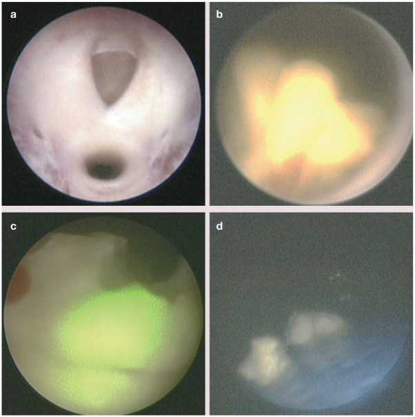
Figure 6.
(a) Cystoscopic appearance of the urethral orifice (top) in a 10-year-old, spayed female domestic shorthair cat. Cystoscopy is performed in dorsal recumbency. (b) Calcium oxalate urocystolith located at the trigone. (c) Laser lithotripsy is performed using a Ho:YAG laser by passing a fiber through the operating port of the cystoscope. The green light is the aiming beam as Ho:YAG laser energy is outside of the visible spectrum. (d) Urocystolith fragments are retrieved using retrieval devices and/or voiding urohydropropulsion

