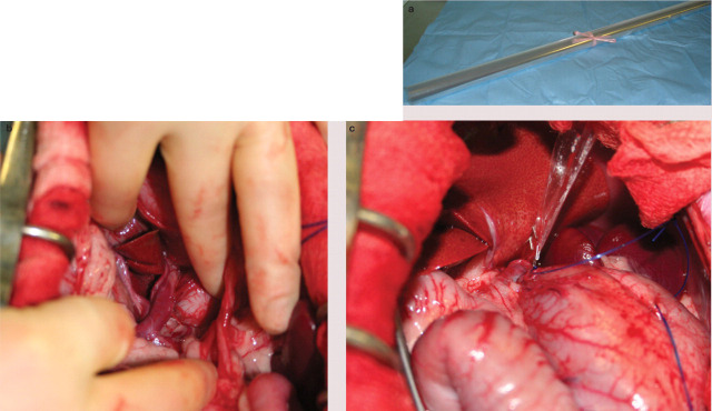
FIG 3.
(a) Roll of (florist's) cellophane for attenuation of a CPSS. The cellophane is cut and folded to form a three layer strip approximately 10 cm long and 4 mm wide. 28 (b) Intraoperative image of a large left gastric shunt in a cat. (c) The shunt has been dissected and a cellophane band has been placed around the vessel. The cellophane has been secured with two haemoclips. The cellophane is cut short prior to abdominal closure. Images (b) and (c) courtesy of Jane Ladlow

