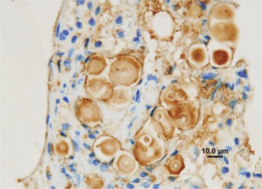
Figure 9.

Use of immunohistology to demonstrate C gattii in histological sections. It is possible to conclusively identify Cryptococcus species in paraffin-embedded formalin-fixed tissue sections using monoclonal antibodies directed against different capsular epitopes. These show up as brown precipitates, highlighting both the yeast cell body and its capsule. Note also the narrow neck budding. Courtesy of Mark Krockenberger
