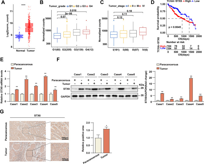
Fig. 1.
STX6 is upregulated in HCC patients and correlates to HCC progression. (A) Results of STX6 expression level analysis in human HCC tissues (n = 347) and normal tissues (n = 50) in TCGA databases. (B) STX6 expression levels in HCC samples of different histologic grades in TCGA databases. Histologic grading grade I, n = 60; grade II, n = 205; grade III, n = 139; grade IV, n = 12. (C) STX6 mRNA levels in HCC samples with different TNM stages in TCGA databases. Stage I, n = 191; Stage II, n = 98; Stage III, n = 97; Stage IV, n = 6. (D) Kaplan-Meier overall survival curves of HCC patients with high and low STX6 expression in the TCGA database. (E) STX6 mRNA levels in paired HCC and paracancerous tissues. β-actin was used to normalize. Data was given as mean ± SD. (F) The WB and quantification results of STX6 in HCC tissues and paracancerous tissues. GAPDH served as the loading control. Data was given as mean ± SD. (G) Results of IHC analysis of STX6 in HCC tissues and paracancerous tissues. Data was given as mean ± SD. For statistical analysis, two-tailed Student’s t-test was used in E-G. * P < 0.05; ** P < 0.01

