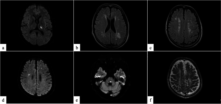
Fig. 2.
Magnetic resonance imaging. a Diffusion-weighted scan and (b, c) fluid-attenuated inversion recovery (FLAIR): Images revealed a new left internal capsule infarction and multiple asymptomatic brain infarctions at the age of 42 years. d, e Diffusion-weighted scan and (f) T2-weighted scan: the last images of consecutive episodes at the age of 48 years; new infarctions in the bilateral cerebral hemispheres and left pons were detected. There were wide-spread periventricular high-intensity lesions in the parietal cerebral area with different time phases

