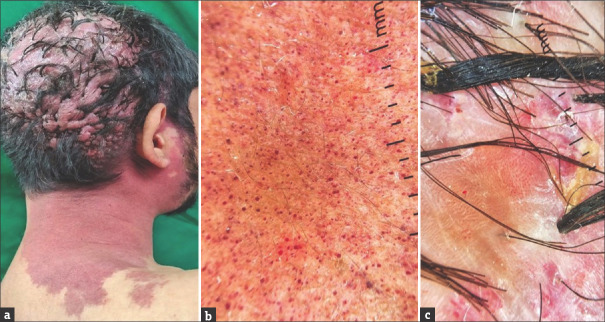
Figure 1.
(a) Port-wine stain extending from the right side of the parieto-occipital region to the side of the neck with multiple hypertrophic, flesh-coloured soft nodules coalescing to form a cerebriform appearance on the parieto-occipital region. (b) Dermoscopy from PWS patch showed red dotted and globular vessels (DermLite DL3N, dry, contact, polarized, x10). (c) Dermoscopy from cerebriform plaque showed red dotted and globular vessels along with white and red structureless areas, tufted hairs and yellowish crusting (DermLite DL3N, dry, contact, polarized, x10)

