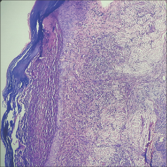
Figure 5.

H and E stain on 100x showing epidermis having hyperkeratosis, parakeratosis, and subcorneal collection of neutrophils (brown arrow). Dermis reveals mixed inflammatory infiltration around blood vessels and hair follicles. (Black arrow). Inflammation extends up to the deep dermis
