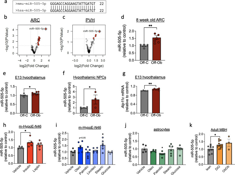
Fig 2. Hypothalamic miR-505-5p expression is increased in offspring of obese mothers from fetal to adult life and in vitro in neurons by exposure to metabolic hormones and fatty acids.
(a) Sequence overlap of mature miR-505-5p in mice and humans. (b, c) Volcano plot of small RNA sequencing results showing significant overexpression of miR-505-5p (red dot surrounded by blue diamond) in the ARC (b) and PVH (c) of 8-week-old offspring born to obese mothers compared to offspring of lean mothers. (d–f) qPCR analysis of miR-505-5p expression in (d) ARC at 8 weeks of age; (e) in whole hypothalamus at E13; (f) in hypothalamic neural progenitor cells extracted at E13 and cultivated as neurospheres for 8 days in vitro. (g) Atp11c (miR-505-5p host gene) mRNA levels in E13 whole hypothalamus. (h) miR-505-5p relative expression of mHypoE-N46 cells exposed to insulin or leptin. (i) miR-505-5p relative expression of mHypoE-N46 cells exposed to oleic acid, palmitic acid, stearic acid, linoleic acid, and glucose for 24 h. (j) miR-505-5p relative expression of C8-D1A (astrocyte) cells exposed to oleic acid, palmitic acid, stearic acid, and glucose for 24 h. (k) miR-505-5p relative expression in MBH in lean, diet-induced obese or genetically obese (Ob/Ob) male mice.* P < 0.05, **P < 0.01. Statistical significance was determined with unpaired t test. Outliers were excluded from (g) for obese group (outlier excluded value 0.878) and (j) for oleic acid treatment (outlier excluded value 1.554). The underlying data are provided as S1 Data file. ARC, arcuate nucleus of hypothalamus; DIO, diet-induced obese; MBH, mediobasal hypothalamus; NPC, neural progenitor cell; PVH, paraventricular nucleus of hypothalamus.

