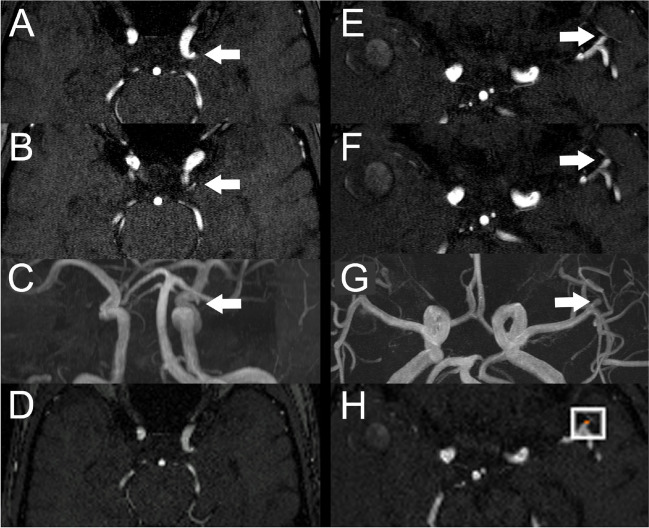
Fig. 2.
A–D Example of a false positive finding reported by the neuroradiologist. A, B Axial TOF image with infundibular origin of a small artery from the left ICA (arrows) reported as an aneurysm by the neuroradiologist in the reading without AI but reported as negative in the reading with AI. C MIP image of the same study with the infundibular origin highlighted (arrow). D Axial reconstruction of TOF-MRA created by the algorithm with no aneurysm detected. E–G Example of a left MCA aneurysm missed by the resident in the reading without AI but correctly reported in the reading with AI. E, F Axial TOF images with small left MCA aneurysm (arrows). G MIP image of the same study with the left MCA aneurysm highlighted (arrow). H Axial reconstruction of the TOF images created by the software with the aneurysm highlighted by a white box

