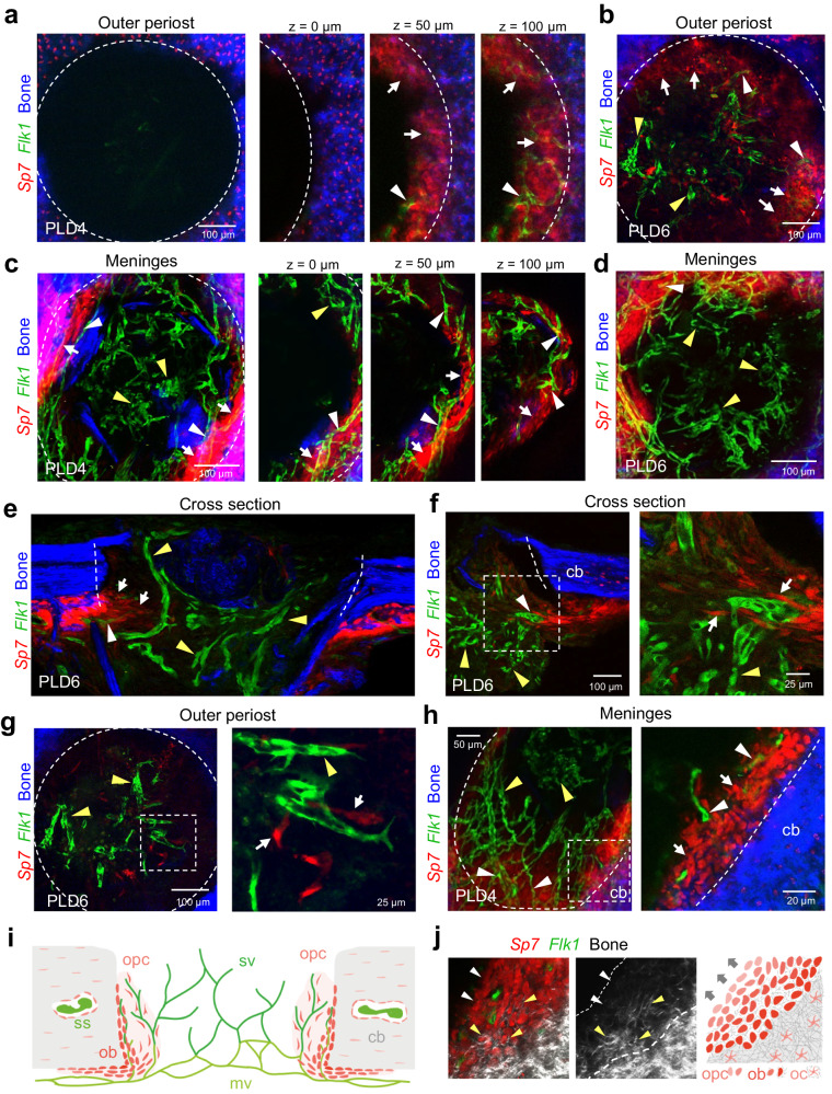
Fig. 4. Osteoprogenitors emerge from the periosteum and collectively colonize the calvarial bone lesion, while blood vessel vascularizes the entire lesion.
a, b Multiphoton imaging of wholemount calvarial bone at PLD4 (a) and PLD6 (b) from the outer periosteal side. a Sp7-mCherry+ (red) osteoblastic cells colonize the injured SHG+ (blue) calvarial bone. Osteoprogenitors are not found at the outer bone edge at PLD4 (z = 0 µm). Deeper layers ( ≥ 50 µm) show early bone-lining osteoprogenitors (white arrows) and associated Flk1+-GFP microvessels (white arrowheads). b The majority of Sp7-mCherry+ osteoprogenitors line the injured calvarial bone (white arrows). Most Flk1+-GFP microvessels in the lesion center are not associated with osteoprogenitors (yellow arrowheads). c, d Multiphoton imaging of wholemount calvarial bone at PLD4 (c) and PLD6 (d) from the inner meningeal side. Early Sp7-mCherry+ osteoprogenitors (white arrows) line the injured bone close to blood vessels (white arrowheads). Microvessels in the lesion center are not associated with osteoprogenitors (yellow arrowheads). e–g Cross sections (e, f) and intravital micrograph (g) of calvarial bone at PLD6 showing early Flk1-GFP+ microvessels and invading, spindle-shaped Sp7-mCherry+ osteoprogenitors. Osteoprogenitors (white arrows) originating from the periosteal layer collectively invade the lesion close to the calvarial bone. Note microvessels with associated osteoprogenitors (white arrowheads), while microvessels in the lesion center are frequently solitary (yellow arrowheads). h Multiphoton imaging showing the meningeal calvarial bone side at PLD4. Flk1+-GFP microvessels (white arrowheads) near the injured calvarial bone are surrounded by Sp7-mCherry+ osteoprogenitors (white arrows). In the lesion center, vessels are not associated with osteoprogenitors (yellow arrowheads). i Schematic showing calvarial bone lesion at PLD6 with sprouting vessels (sv) originating from meningeal vessels (mv) and invading osteoprogenitor (opc). Osteoprogenitors remain close to the injured calvarial bone, while sprouting vessels penetrate and vascularize the entire lesion. Area of angiogenic-osteogenic coupling is shown in light red. Microvessels in the center of the lesion are devoid of osteoprogenitors. ss: sinusoidal vessels, ob: osteoblasts, oc: osteocytes. j Multiphoton imaging showing the growing calvarial bone edge and corresponding schematic. Sp7-mCherry+ osteoprogenitors invade the bone lesion as a multicell layer and gradually differentiate to matrix forming osteoblasts. Grey arrow indicates direction of bone growth. Reproducibility was ensured by n = 3 or more biologically independent experiments.

