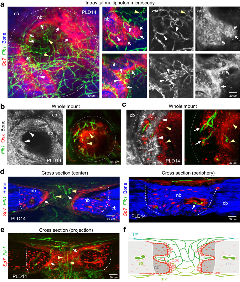
Fig. 5. Osteoblasts lining the growing bone collectively invade the vascularized calvarial bone lesion.
a Intravital multiphoton microscopy showing the expanding SHG+ (blue) calvarial bone lined by a multicellular layer of Sp7-mCherry+ (red) osteoblastic cells and Flk1-GFP+ (green) microvessels vascularizing the calvarial bone lesion in Flk1-GFP, Sp7-mCherry double transgenic mice at PLD14. Maximum intensity projection (left). Zoom-in views (right) show association of microvessels (white arrowheads) with a dense layer of osteoblastic cells at the front of the growing bone (nb) (white arrows). Note that microvessels in the uncalcified lesion center are not associated with osteoprogenitors (yellow arrowheads). b, c Multiphoton microscopy of whole mount calvarial bone at PLD14. b Maximum projection shows Osx+ (red) osteoblasts and progenitors, Flk1-GFP+ (green) microvessels, SHG+ (white) calvarial bone (cb) and new bone (nb) in Flk1-GFP+ transgenic mice stained with anti-Osx antibodies. c Zoom-in views show single planes. White arrowheads point to the front of growing SHG+ bone matrix (nb). Yellow arrow heads indicate the front of a multicellular layer of Osx+ osteoblastic cells that precedes the SHG+ bone front. White arrows point to microvessels enclosed in the newly formed bone in close proximity to Osx+ osteoblastic cells. d Cross sections showing Flk1-GFP+ (green) microvasculature, Sp7-mCherry+ (red) osteoblasts and progenitors lining the SHG+ (blue) growing bone, osteocytes enclosed by SHG+ new bone (nb) at PLD14. Two single planes (left: center, right: periphery) through the lesion are shown. White arrows (right) indicate the multicellular layer of osteoblasts and progenitors lining the new bone. Yellow arrowheads indicate microvessels. Arrow (left) indicates an early bone marrow cavity. e Maximum intensity projection of a cross section showing Flk1-GFP+ (green) microvasculature, Sp7-mCherry+ (red) osteoblasts and progenitors at PLD14. White arrows indicate osteoblasts at the front of the growing bone. Yellow arrows indicate microvessels connecting meningeal and periosteal vessels in the uncalcified lesion. f Schematic cross section of calvarial bone at PLD14. Meningeal vessels (mv) form a vascular network within the lesion that connects to outer periosteal vessels (pv). Osteoblastic cells expand the growing bone into the vascularized lesion area. Microvessels in the uncalcified lesion are devoid of osteoprogenitors. cb: calvarial bone, oc: osteocytes ob: osteoblasts, opc: osteoprogenitors, ss: sinusoidal capillaries. Reproducibility was ensured by n = 3 or more biologically independent experiments.

