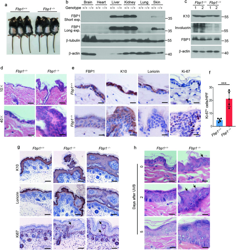
Fig. 3. FBP1 is essential for epidermal homeostasis and response to UVB irradiation.
a Photos of 6-week-old Fbp1+/+ and Fbp1−/+ mice. b Western blot analysis of the expression of FBP1, β-tubulin and β-actin in indicated organs obtained from Fbp1+/+ and Fbp1−/+ mice. Exp exposure. c Western blot analysis of the expression of K10, involucrin, FBP1 and β-actin in skin tissue from Fbp1+/+ and Fbp1−/+ mice. d Representative H&E staining of skin from 6-week-old Fbp1+/+ and Fbp1−/+ mice. Scale bars in upper images: 100 μm, scale bars in lower images: 25 μm. e Immunohistochemical staining for FBP1, K10, loricrin and Ki-67 of skin from 6-week-old Fbp1+/+ and Fbp1−/+ mice. Scale bars: 25 μm. f The quantification of Ki-67+ keratinocytes in skin sections from 6-week-old Fbp1+/+ and Fbp1−/+ mice, n = 3 mice per genotype. HPF high-power field. g Immunohistochemical staining for K10, loricrin and Ki-67 of skin from neonatal Fbp1+/+, Fbp1−/+ and Fbp1−/− mice. Scale bars: 100 μm. Arrows indicated protruding Ki-67+ basal keratinocytes. h Representative H&E staining of skin from 6-week-old Fbp1+/+ and Fbp1−/+ mice collected at indicated days after irradiation. Scale bars: 50 μm. Arrows indicated retained cell nuclei in the cornified layer. Data are shown as mean ± s.d. Statistical analyses in (f) were performed with Student’s t-tests. ***p < 0.001.

