Figure 1.
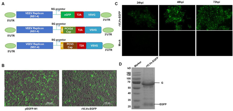
Construction and identification of rVLVs-EGFP. (A) Diagram of VLV DNA constructs used to generate rVLVs expressing EGFP, PCV2d Cap, and PCV3 Cap proteins. Sequences encoding the EGFP, PCV2d Cap, and PCV3 Cap proteins are inserted under the viral subgenomic RNA promoter upstream of a T2A ribosomal skipping site and the VSV G protein. (B) Fluorogram of BHK-21 cells transfected with rVLVs-EGFP mRNA at 72 hpt post-transfection. A transfection of the pEGFP-N1 plasmid was used as a positive control. (C) Fluorogram of rVLVs-EGFP infected BHK-21 cells. BHK-21 cells were infected with supernatants harvested after transfection of BHK-21 cells with rVLVs-EGFP genomic RNA at 72 hpt. At the indicated time points, the expression of the RABV-G protein was analyzed by the accumulation of green fluorescence. (D) SDS-PAGE analysis of supernatants from BHK-21 cells infected with rVLVs-EGFP. The rVLVs-EGFP was separated by a 12% SDS-PAGE gel, and the protein bands were observed by coomassie staining. The position and molecular weight of the rVLVs-EGFP structural protein VSV-G were also indicated.

