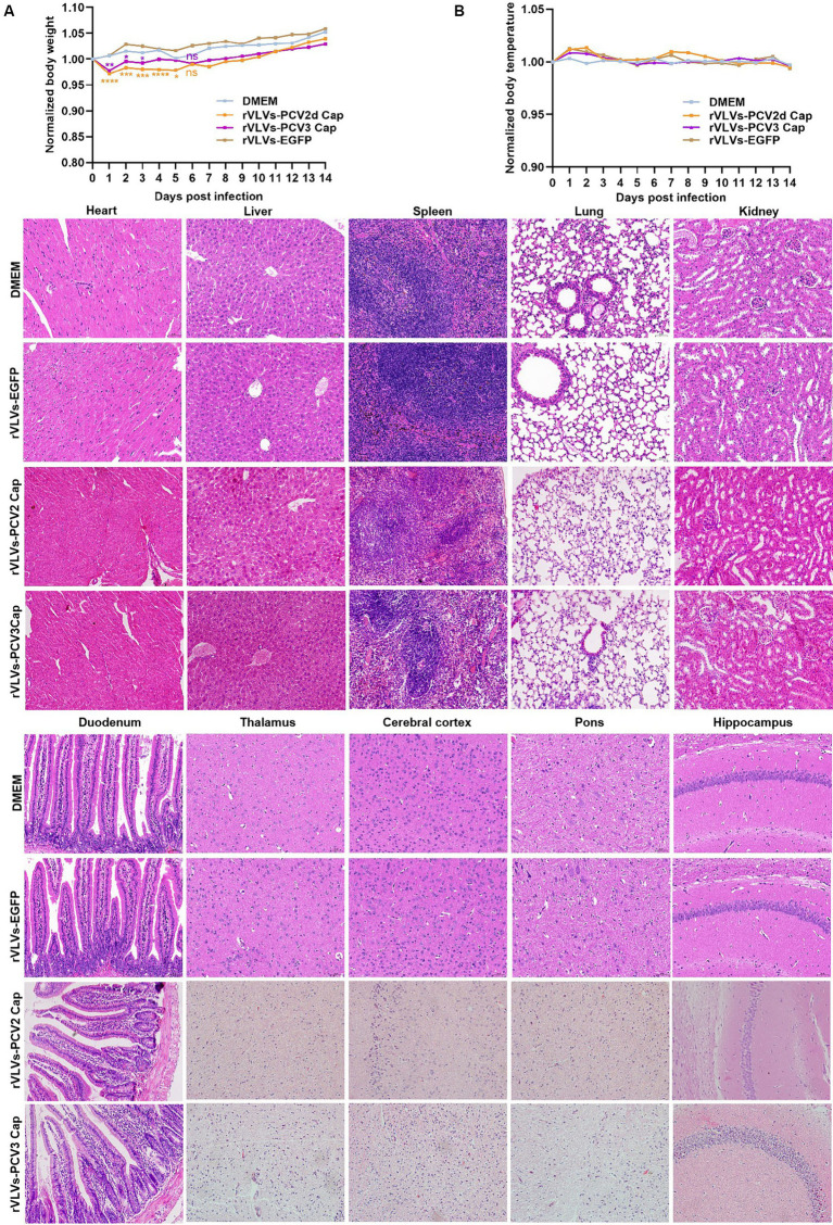
Figure 4.
Virulence of rVLVs in mice. (A,B) Groups of 6-week-old male C57 mice (n = 7/group) were via i.m. infected with rVLVs-EGFP, rVLVs-PCV2d Cap, and rVLVs-PCV3 Cap 105 FFU or mock-infected with 100 μL DMEM. (A) Body weight loss was monitored daily for 14 days, and (B) body temperature was monitored daily for 14 days. (C) Groups of 6-week-old male C57 mice (n = 7/group) were i.m. injected with rVLVs-EGFP, rVLVs-PCV2d Cap, and rVLVs-PCV3 Cap 105FFU or mock-infected with 100 μL DMEM. At 7 d.p.i., all groups of mice were euthanized, parenchymal organs such as the heart, liver, spleen, lungs, kidneys, and brain and as well as the hollow organ duodenum were taken to make paraffin tissue sections for HE staining, and histopathological analysis. Sagittal sections of the brain were H&E stained and histopathologically analyzed in different brain regions including the pons, thalamus, hippocampus, cerebral cortex, and presented representative histological changes. Data are presented as means ± SD. Asterisks indicate significant differences between the indicated experimental groups, *p < 0.05, **p < 0.01, ***p < 0.001 (One-way ANOVA). Orange asterisks represent the difference between rVLVs-PCV2d Cap and DMEM and Purple asterisks represent the difference between rVLVs-PCV3 Cap and DMEM.

