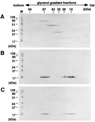
FIG. 5.
Sedimentation profiles of myristoylated and nonmyristoylated MA domains. Purified MA proteins were layered onto 15 to 30% glycerol gradients and centrifuged at 48,000 rpm for 40 h. Fractions from the bottom to the top (left to right) were subjected to SDS-PAGE, and proteins were detected by Western blotting using an antipolyhistidine monoclonal antibody (Sigma). (A) Low-molecular-weight calibration markers for sedimentation (Amersham Pharmacia Biotech) stained with Coomassie brilliant blue; (B) wild-type MA; (C) nonmyristoylated MA(G2A), detected by Western blotting. Lane M shows prestained molecular weight markers (Bio-Rad) for SDS-PAGE.

