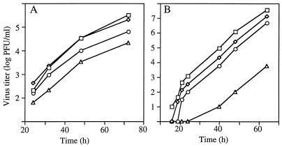
FIG. 5.
Virus titers of VPQAQA variants in vertebrate cells. (A) Vero cells (105) in 24-well dishes were infected (MOI, ∼0.1) with YFV 17D (squares) or VPQAQA variants V2(P117→L) (diamonds), V4(H103→L) (circles), and V7(T107→I) (triangles), and samples were taken between 24 and 72 h p.i. for assay of infectivity titers. (B) BHK cells were electroporated with full-length RNA transcripts (∼0.1 μg) incorporating the coding regions for the C-prM junctions of V2(P117→L), V4(H103→L), V7(T107→I), and YFV 17D. Electroporated cells (2 × 10⁶) were plated in 60-mm dishes, and supernatant samples were taken between 16 and 64 h posttransfection for assay of infectivity titers. Data for 17D RNA (squares), V2′(P117→L) RNA (diamonds), V4′(H103→L) RNA (circles), and V7′(T107→I) RNA (triangles) are shown.

