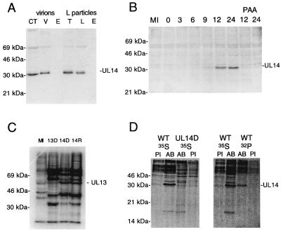
FIG. 3.
Immunological identification of the UL14 protein and assessment of UL13 expression. (A) HSV-1 virions (V) or L particles (L) and fractions derived therefrom (CT, capsid-tegument; T, tegument; E, envelope) were separated on a 10% polyacrylamide gel, transferred to a Hybond ECL membrane, and immunoblotted with the UL14 antiserum. Positions of the UL14 protein and marker proteins are indicated. (B) HSV-1-infected cells harvested at various times postinfection were separated on a 10% polyacrylamide gel, transferred to a Hybond ECL membrane, and immunoblotted with the UL14 antiserum. Positions of the UL14 protein and marker proteins are indicated. The mock-infected (MI) sample was harvested at 24 h. Two samples were maintained in the presence of phosphonoacetic acid and harvested at 12 and 24 h postinfection. (C) In vitro-phosphorylated nuclear extracts from mock-infected cells (MI) or cells infected with a UL13 null mutant (13D [11]), UL14D (14D), or UL14R (14R) were separated on a 10% polyacrylamide gel and autoradiographed. Positions of the labeled UL13 protein and marker proteins are indicated. (D) 35S- or 32P-labeled proteins from cells infected with wild-type HSV-1 (WT) or UL14D were immunoprecipitated with preimmune serum (PI) or the UL14 antiserum (AB), separated on 15% polyacrylamide gels, and phosphorimaged. Positions of the UL14 protein and marker proteins are indicated.

