Figure 2.

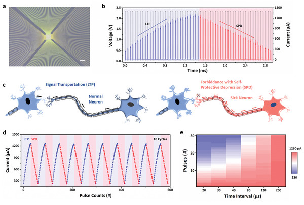
a) The scanning electron microscopy images of the Au/PBFCL10/Ag memristive device. Bar scale: 100 µm. b) The LTP and SPD effects of the Au/PBFCL10/Ag device with continuous 59 pulses (amplitude: 2.3 V, width: 10 µs, and interval: 40 µs). c) The schematic diagram of the signal transportation in normal neuron and signal forbiddance in sick neuron by post‐synapse, corresponding to LTP and SPD effects in Figure 2b, respectively. d) The self‐oscillated behavior of the Au/PBFCL10/Ag synaptic device. e) The variation amplitude of the current under the pulse stimulation with different time interval.
