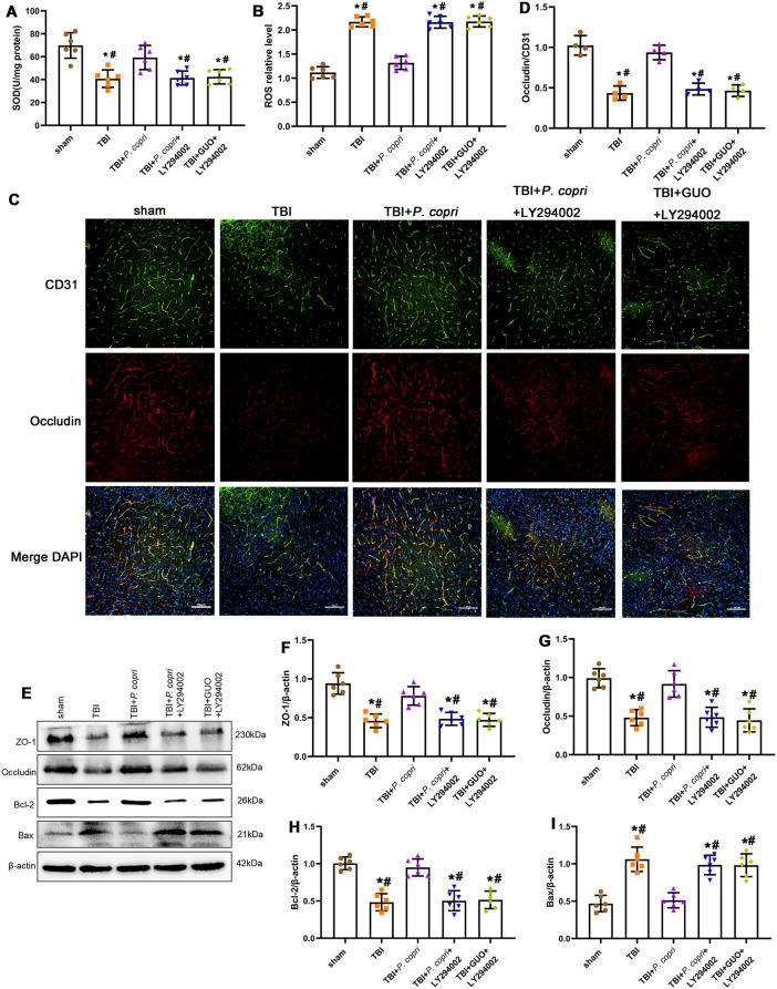
Fig. 12.
The GUO-PI3K/Akt axis was involved in the effects of P. copri on oxidative stress, blood-brain barrier and neuronal apoptosis in TBI mice. A-B. Oxidative stress after TBI. SOD activity (A) and ROS level (B). *, p < 0.05 vs. Sham; #, p < 0.05 vs. TBI + P. copri, n = 6 per group. C. Representative images of double immunofluorescence staining for Occludin and CD31, nuclei were stained with DAPI (blue). Scale bar = 100 μm. D. Quantitative analysis of relative Occludin fluorescence intensity in different groups. *, p < 0.05 vs. sham; #, p < 0.05 vs. TBI + P. copri, n = 4 per group. E. Representative Western blot bands of ZO-1, Occludin, Bcl-2, Bax and β-Actin at the lesion sites after TBI. F-I. Quantitative analysis of the relative density of ZO-1 (F), Occludin (G), Bcl-2 (H) and Bax (I). *, p < 0.05 vs. sham; #, p < 0.05 vs. TBI + P. copri, n = 6 per group. One-way ANOVA was used to compare means of different groups followed by a Tukey post hoc multiple-comparisons test

