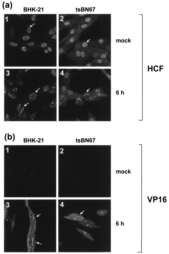
FIG. 6.
Intracellular distribution of HCF and VP16 in BHK-21 and tsBN67 cells infected at 39.5°C. BHK-21 and tsBN67 cells grown for 2 days at 39.5°C were mock infected or infected with HSV-1 at an MOI of 10 and fixed 6 h later. Indirect immunofluorescence was carried out with the anti-HCF polyclonal antibody (a) or LP1, the anti-VP16 MAb (b).

