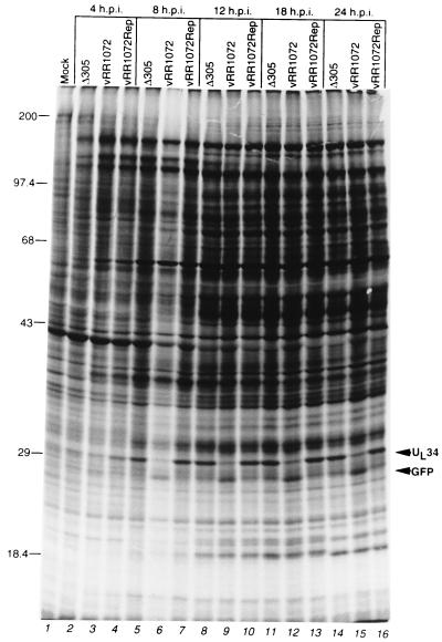
FIG. 6.
Protein synthesis in Vero cells infected with wild-type, UL34-negative, and repair viruses. Shown is an autoradiographic image of SDS-PAGE-separated proteins from Vero cells either mock infected (lane 1) or infected with the indicated virus for 4 h (lanes 2 to 5), 8 h (lanes 6 to 8), 12 h (lanes 9 to 11), 18 h (lanes 12 to 14), or 24 h (lanes 14 to 16) and labeled with [35S]methionine for 2 h prior to harvest. The migration positions of size standards (in kilodaltons) are indicated at the left. The migration positions of UL34 and GFP are indicated by arrowheads at the right. The overall lower level of label seen in lane 6 is due to a corresponding overall lower level of protein loaded in that lane. p.i., postinfection.

