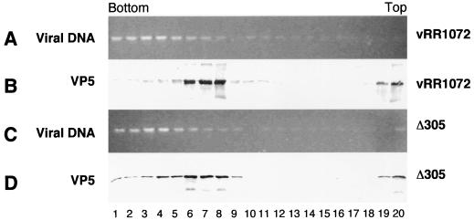
FIG. 8.
Nuclear capsids and encapsidated DNA in wild-type and UL34-negative virus. Photographic images show electrophoretically separated and ethidium bromide-stained DNA (A and C) and Western-blotted proteins (B and D) from sucrose gradient fractionation of DNase I-treated nuclear lysates from cells infected with either vRR1072 (A and B) or Δ305 (C and D).

