FIG. 9.
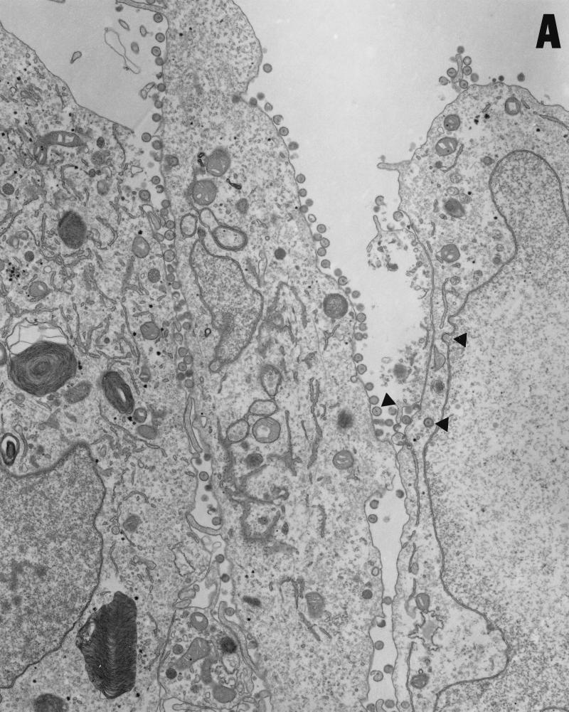
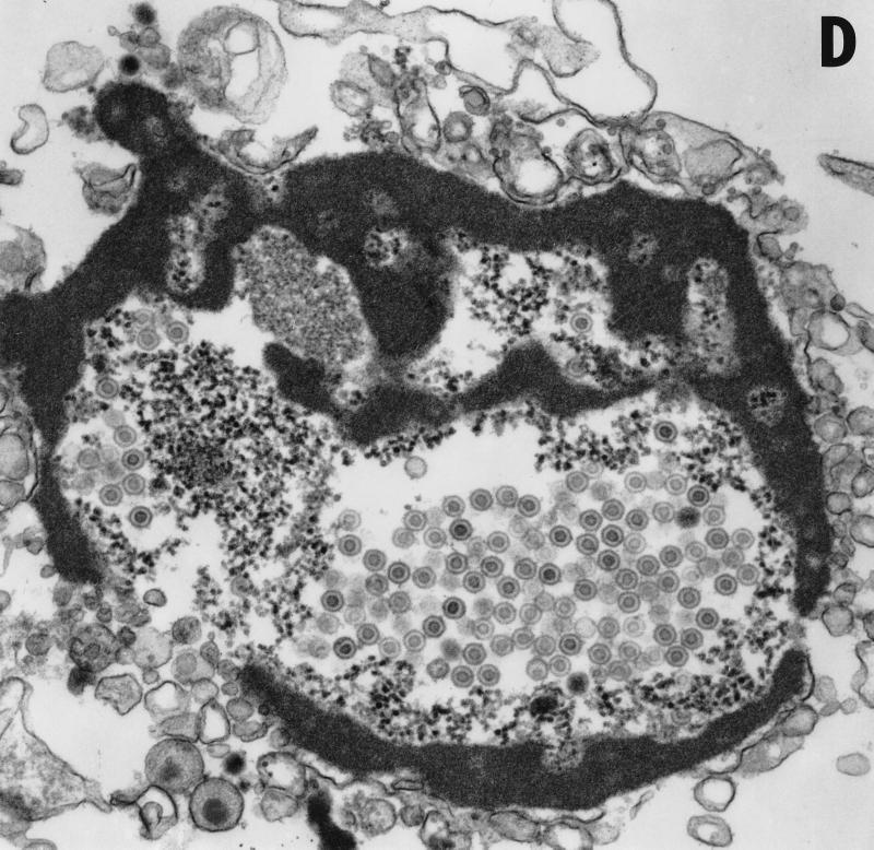
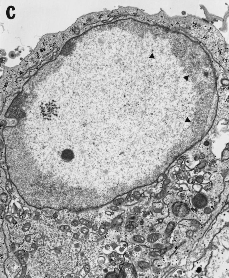
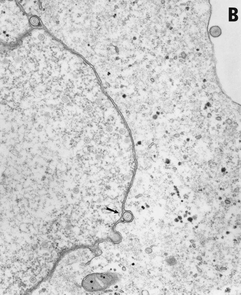
Transmission EM analysis of cells infected with wild-type and UL34-negative virus. Micrographs show Vero cells infected with either HSV-1(F) (panels A and B) or vRR1072(TK+) (panels C to F) for 20 h. (A) Wild-type-infected Vero cells showing envelopment of capsid at the inner nuclear membrane, enveloped virus particles in vesicles in the cytoplasm, and enveloped virus at the surface of, and between cells. Examples of each are indicated by arrowheads. Final magnification is ×13,500. (B) Wild-type-infected Vero cell showing capsids in the nucleus and enveloped virus particles between the inner and outer nuclear membranes (one example indicated by arrow). Final magnification is ×40,500. (C) Deletion mutant-infected Vero cell showing capsids in the nucleus (a few examples indicated with arrowheads) but no cytoplasmic or cell-surface-associated enveloped virus particles. Final magnification is ×13,500. (D) Cell fragment from deletion mutant-infected Vero cells containing numerous viral capsids. Final magnification is ×40,500.


