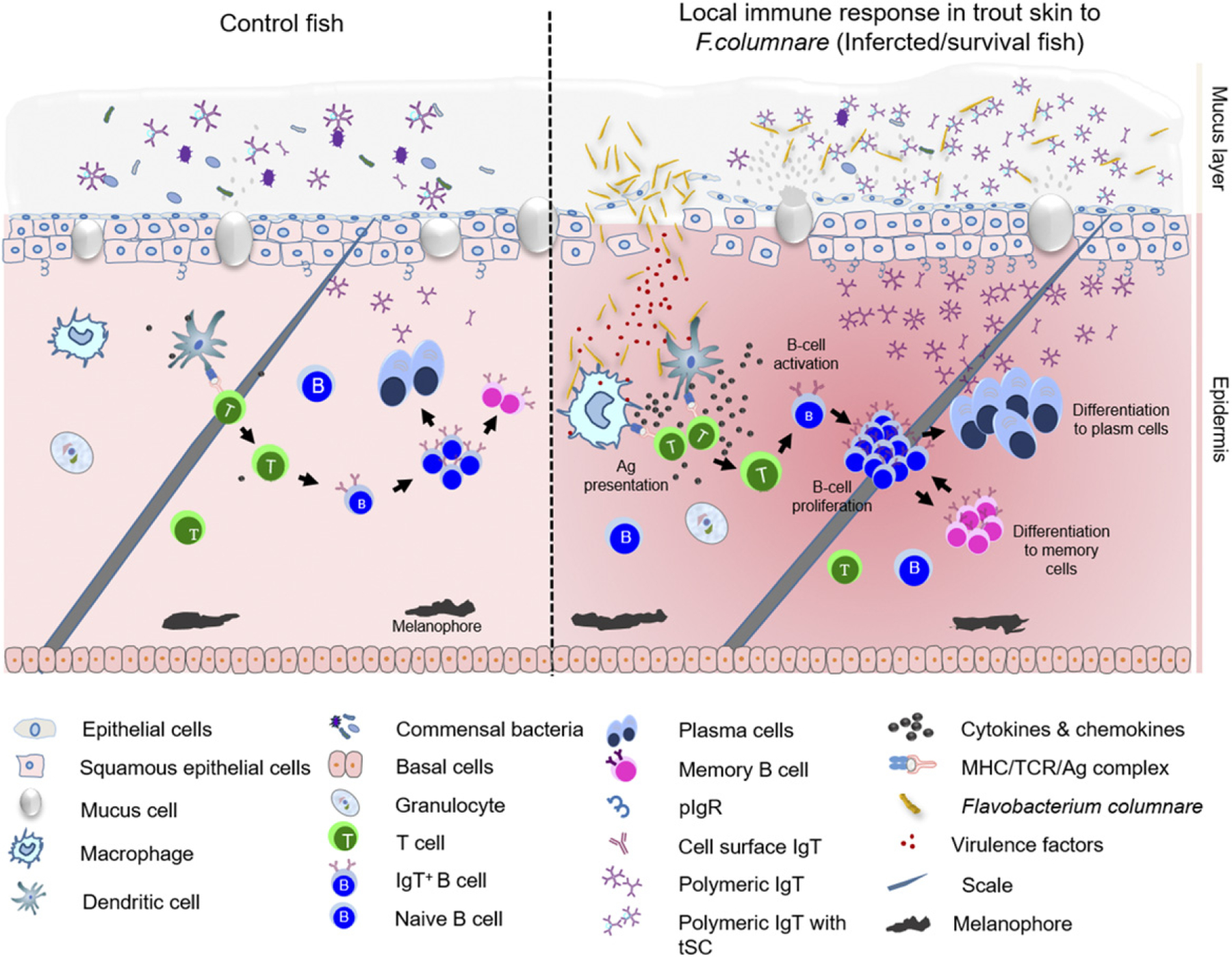
FIGURE 8.

Proposed model of local IgT and IgT+ B cell induction in the skin after F. columnare infection. The presumed structure of skin displaying mucus layer, epithelial layer, and dermis. The proposed model contains two partitions: control (left) and infected/survivor (right). Induction of local IgT responses in the trout skin based on our findings. On the left is the scheme of a typical skin structure in control (naive) teleost fish. The number of IgT+ B cells in control fish skin is low. IgT are produced by IgT-secreting B cells and transported from the epithelium into the skin mucus layer via pIgR. The sIgT coats the majority of microbiota in the skin surface. In the skin epidermis, abundant mucus cells are present, and their production contains antibacterial molecules to protect host against pathogens. Upon F. columnare invasion, their Ag can be taken up by APCs and presented to naive CD4+ T cells. IgT+ B cells are then activated by Ag-specific CD4+ T cells and start to proliferate locally and differentiate into plasma cells in skin epithelial layer. These differentiated IgT-secreting cells can produce large amounts of pathogen-specific IgT, which can be transported by pIgR into skin mucus layer, where those pathogen-specific IgT can specifically bind to the bacteria F. columnare. Alternatively, some IgT+ plasma cells may differentiate further into memory IgT+ B cells. When F. columnare invade the host again, the memory IgT+ B cells would directly proliferate and differentiate into plasma cells and then rapidly produce specific IgT to binding F. columnare.
