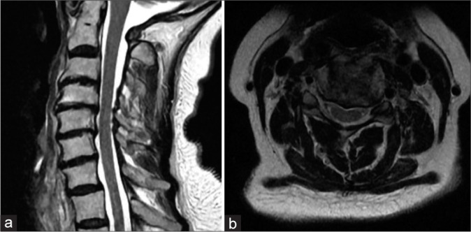
Figure 2:

(a) Sagittal T2-weighted magnetic resonance imaging (MRI) of the cervical spine showing multilevel spondylotic changes. (b) Axial T2-weighted MRI of the cervical spine at the C4–5 level showing severe left and moderate right foraminal stenosis.
