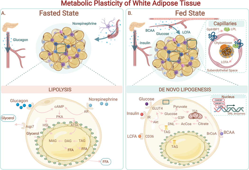
Figure 3. Metabolic Plasticity of White Adipose Tissue.
(A) Fasted state. Adipocytes release free fatty acids (FFAs) and glycerol via lipolysis in response to external stimulation (i.e., norepinephrine, glucagon). Binding of norepinephrine to the adrenergic receptor (AR) on adipocytes drives the elevation of cAMP and PKA activation. PKA stimulates the hydrolysis of triglycerides (TAG), diacylglycerol (DAG), and subsequently monoacylglycerol (MAG) through activation of the endogenous lipases ATGL and HSL. FFAs and glycerol are secreted into the systemic circulation to supply fuel to other tissues.(B) Fed State. Adipocytes have access to multiple sources of circulating nutrients, including: 1) Long Chain Fatty Acids (LCFA) from Very Low-Density Lipoprotein (VLDL) (LPL-mediated hydrolysis of triacylglycerols from VLDL in capillaries to generate FFAs); 2) glucose; 3) branched-chain amino acids (BCAA). De novo lipogenesis (DNL) uses Acetyl-CoA (AcCoA) as the primary building block for fatty acid synthesis. Synthesized fatty acids are esterified into triglycerides (TAG) and stored in lipid droplets. Expression of enzymes involved in DNL (i.e., fatty acid synthase (FAS); acetyl-CoA carboxylase (ACC)) are positively regulated by hormones (i.e., insulin) and by transcription factors such as Carbohydrate response element binding protein (ChREBP), Liver X receptor alpha (LXRa) and Sterol response element binding protein 1c (SREBP1c). TAGs stored in the lipid droplet are released by lipolysis during periods of energy demand.

