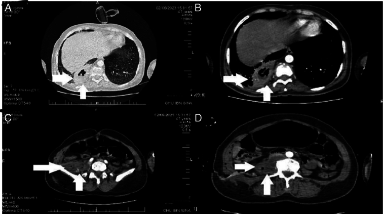
Figure 2.

Postoperative CT scan imaging showing a psoas abscess, with right lung fistulization:. A. Parenchymal window of a cross-sectional image of thoracic CT illustrating fistulization of psoas abscess into pulmonary parenchyma. B. Mediastinal window of a cross-sectional image of thoracic CT illustrating fistulization of psoas abscess into pulmonary parenchyma. C. Pelvic CT scan revealing a right paravertebral hypodense collection measuring 75 mm in its long axis, accompanied by pubic osteitis. D. Abdominal CT scan demonstrating a right paravertebral hypodense collection containing air bubbles, with enhanced wall after contrast agent injection.
