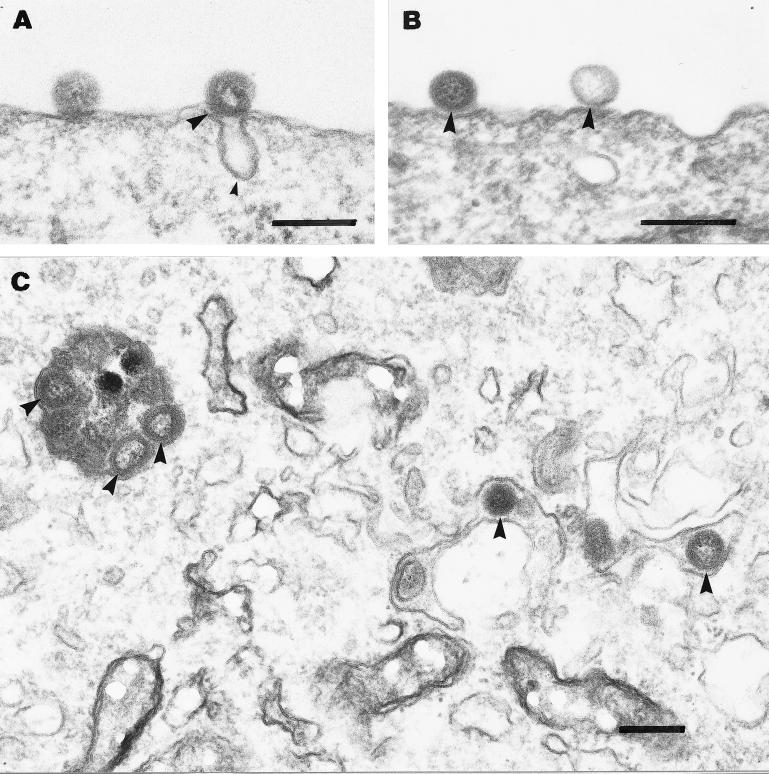
FIG. 5.
Electron micrographs of the early steps in ISAV infection. (A and B) Association of ISAV (big arrowheads in all panels) with the plasma membrane after 4 h of binding at 4°C. Virus was found associated with either invaginations presumably representing caveolae (small arrowhead) (A) or flat plasma membrane stretches without any visible coating (B). Further incubation of cells for 4 h with chloroquine (0.1 mM) at 15°C led to intracellular accumulation of ISAV in vesicular and tubular structures (C). Bars, 200 nm.

