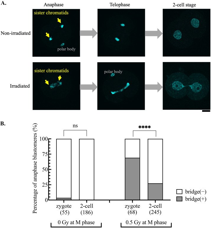
Fig. 1.
Exceptionally high formation rate of anaphase bridges in zygotes irradiated during mitosis. A: Representative images of nonirradiated and 10 Gy-irradiated embryos, which were fixed and stained with DAPI at anaphase and telophase of the first cell cycle and 2 h after entering the 2-cell stage. Scale bar, 20 μm. B: The percentage of anaphase cells (blastomeres) with bridges. Zygotes and 2-cell stage embryos were arrested with nocodazole and irradiated at 15 and 35 HPI, respectively. One hour after irradiation, embryos were moved to a nocodazole-free medium for further development and fixed at anaphase. The cumulative results from three independent experiments are shown. The total number of blastomeres examined for each condition is indicated in the figure. Fisher’s exact test was used for statistical analysis (ns, not significant; **** P < 0.0001).

