Fig. 2.
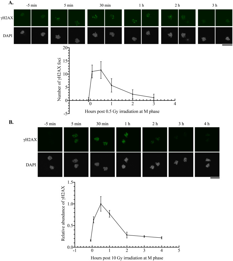
Efficient resolution of γH2AX in irradiated M phase zygotes. A: γH2AX dynamics in zygotes irradiated with 0.5 Gy at 15 HPI (M phase). The upper panel shows representative images at each time point. Images of paternal and maternal chromosomes were taken separately to accurately count the number of γH2AX foci. Three independent experiments were conducted, and more than six embryos were examined for each condition in each experiment. B: γH2AX dynamics in zygotes irradiated with 10 Gy at 15 HPI (M phase). The upper panel shows representative images at each time point. Relative intensity of γH2AX to DAPI was measured and normalized to the average value at 30 min post-irradiation. Four independent experiments were conducted, and more than six embryos were examined for each condition in each experiment. Scale bar, 20 μm. Error bars indicate standard deviations.

