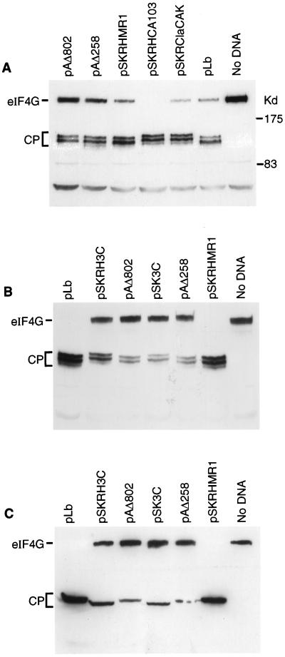
FIG. 4.
FMDV 3C induces cleavage of eIF4G (A). BHK cells were infected with vTF7-3 (as for Fig. 3) and transfected with the indicated plasmids (Fig. 3A). After 20 h, cell extracts were prepared and analyzed by SDS-PAGE (6% polyacrylamide) and immunoblotting with an N-terminal region-specific anti-eIF4G antiserum. (B and C) In a similar experiment vTF7-3-infected BHK cells were transfected with the indicated plasmids, and cell extracts were analyzed by SDS-PAGE (6% polyacrylamide) and immunoblotting with antisera specific for the N-terminal region of eIF4G (B) or the C-terminal fragment of eIF4G (C).

