Figure 4.
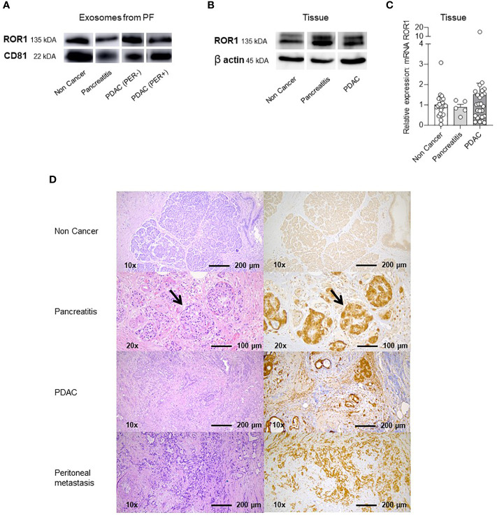
Western Blots (WB), qPCR and Immunhistochemistry (IHC) of exosomes in PF and lysed tissue. (A) Western Blot with ROR1 and CD81 of isolated exosomes from PF. Both proteins are expressed on the exosomes of all different groups (Non Cancer, Pancreatitis, PDAC (PER-), PDAC (PER+). For uncropped WB refer to Supplementary Figure S2 . (B) Western Blot of lysed tissue from Non Cancer, Pancreatitis, PDAC. ROR1 and also β-Actin as loading control is expressed in all three groups. For uncropped WB refer to Supplementary Figure S2 . (C) qPCR analysis and relative ROR1 expression of pancreatic tissue. ROR1 is expressed on NC, CP and PDAC tissue showing no significant differences between the groups but a slightly higher expression in the PDAC tissue. (D) Immunohistochemistry (IHC) and HE staining of non-cancerous exocrine pancreatic tissue, CP, PDAC and peritoneal metastasis. The exocrine pancreas is ROR1 negative. In the CP tissue ROR1 positive islet cells are shown in the higher magnification (arrows). The fibrotic tissue is negative. In PDAC and the peritoneal metastasis the morphologic tumor cells are ROR1 positive.

