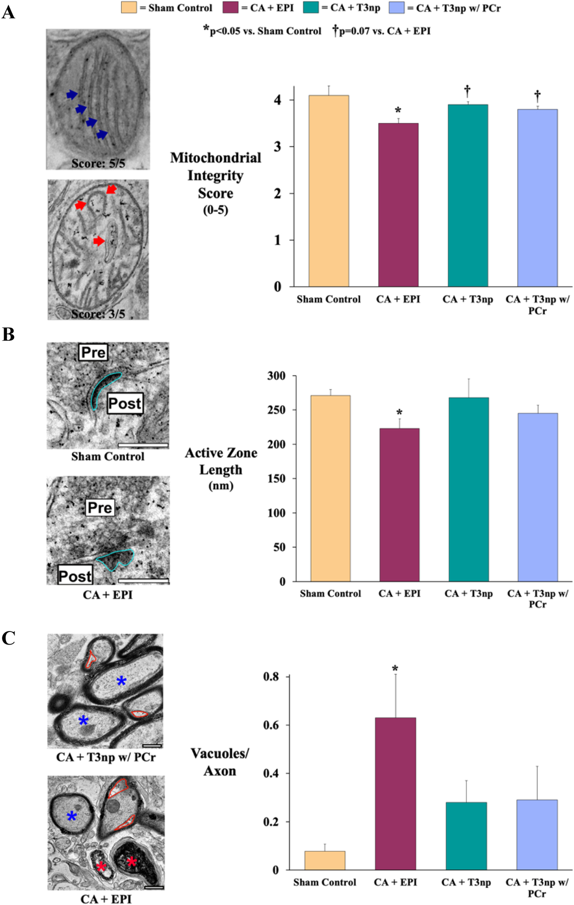
Figure 5: Post-ROSC Hippocampal Ultrastructure in Swine Resuscitated with T3np, T3np+PCR, and EPI After Cardiac Arrest.

(A) Hippocampal mitochondrial integrity was assessed by examining cristae organization and fragmentation (left panel; blue arrows), with assignment of a score on a scale from 1–5 (5 = mitochondria with preserved ultrastructural integrity). Based on this scoring system, mitochondrial integrity was significantly reduced in EPI-treated animals (left panel; red arrows) compared with sham controls but was largely preserved in T3np-treated animals. (B) Active zone length (left panel; outlined in blue) at neuronal synapses was significantly reduced in EPI-treated animals vs. sham controls, suggestive of impaired neurotransmitter release from synaptic vesicles in these animals. However, T3np-treated animals did not exhibit significant changes in active zone length compared with sham controls. (C) Ultrastructural changes to hippocampal axons were assessed as shown in the left panel, with a normal axon labeled with a blue asterisk, dysfunctional axons labeled with red asterisks, and vacuoles outlined in red. Dysfunctional axons were more prevalent in EPI-treated animals and this group also exhibited a significant increase in axonal vacuolization, an early sign of axonal injury. However, this was attenuated in animals treated with either formulation of T3np, indicating better preservation of axonal structure in these groups. Values are mean±SEM. *p<0.05 vs. sham control; †p=0.07 vs. CA+EPI. CA = cardiac arrest; EPI = epinephrine; T3np = triiodothyronine nanoparticles; T3np+PCr = triiodothyronine nanoparticles with encapsulated phosphocreatine.
