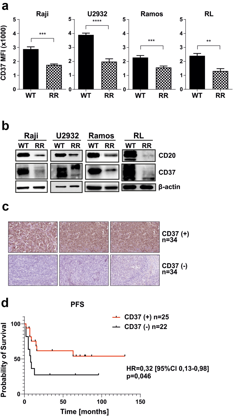
Figure 3.

CD37 is downregulated in RR cells.
(a) WT and RR cells were stained with FITC-conjugated anti-CD37 mAb. Dead cells were discriminated upon staining with PI. The results are presented as MFI of WT and RR cells (mean ± SD). Statistical significance was determined with Welch’s t-test, **p<0.01, ***p<0.001, ****p<0.0001. The experiments were repeated independently four times. (b) The levels of CD20 and CD37 were assessed with Western blotting in whole-cell lysates. β-actin was used as loading control. The experiments were repeated independently three times. (c) CD37 expression was evaluated in 68 primary paraffin-embedded samples by a pathologist blinded to the clinical data of the patients. Representative stainings for CD37-positive and CD37-negative stainings are shown. (d) Kaplan–Meier curves were plotted for PFS in CD37- and CD37+ patients. The survival curves were compared with Peto and Peto’s generalized Wilcoxon test.
