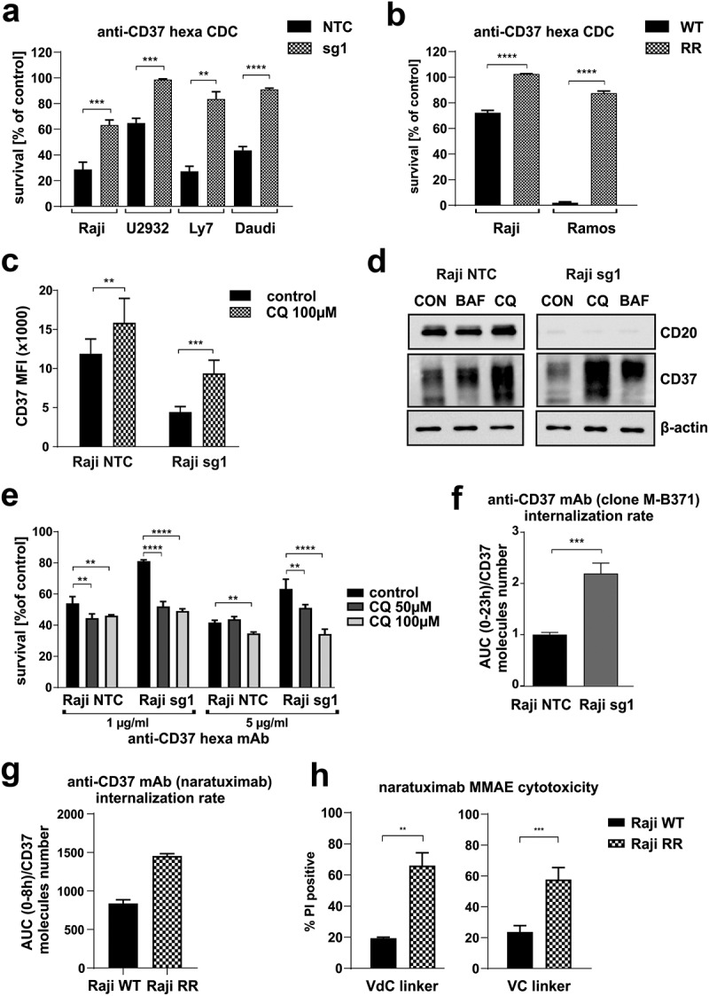
Figure 4.

CD37 decrease leads to impaired efficacy of anti-CD37 mAbs-mediated CDC, which can be restored using lysosome inhibitors.
Equal amounts of NTC and CD20 KO cells (a) and WT and RR Raji cells (b) were incubated for 1 h (Raji, Ramos, Daudi, Ly7) or 4 h (U2932) with 10 µg/ml anti-CD37 mAb and 20% human AB serum as a source of complement. Cell viability was assessed with PI staining. The survival of cells is presented as a percentage of control cells without antibody (mean± SD). Statistical significance was determined using unpaired t-test, ***p<0.001, ****p<0.0001 vs controls. (c) Equal amounts of NTC and CD20 KO cells were incubated for 24 h with 100 µM chloroquine. Thereafter, the cells were stained with PE-conjugated anti-CD37 mAb. The results are presented as MFI of NTC and sg1 cells (mean± SD). Statistical significance was determined using 2-way ANOVA with Sidak’s post hoc test, **p < 0.01, ***p < 0.001 vs controls (d) The levels of CD20 and CD37 were assessed with Western blotting in whole-cell lysates. β-actin was used as loading control. (e) Equal amounts of NTC and CD20 KO Raji cells preincubated for 24 h with 50 or 100 µM chloroquine were treated for 1 h with anti-CD37 mAb (5–10 µg/ml) and 20% human AB serum as a source of complement. Cell viability was assessed with PI staining. The survival of cells is presented as a percentage of control cells without antibody (mean± SD). Statistical significance was determined using 2-way ANOVA with Sidak’s post hoc test, **p<0.01, ****p<0.0001 vs controls. (f) Internalization rate of anti-CD37 mAb (clone M-B371) was determined in Raji NTC and sg1 cells incubated in IncuCyte with equal amounts of mAb conjugated to pH sensitive dye activated in lysosome. Statistical significance was determined using unpaired t-test, ***p<0.001 vs controls. (g) Internalization rate of anti-CD37 mAb (clone K7153A) was determined in Raji WT and RR cells incubated in IncuCyte with equal amounts of mAb conjugated to pH sensitive dye activated in lysosome. The internalization rates are shown as fluorescence AUC normalized to number of CD37 molecules per cell. (h) Equal amounts of Raji WT and RR cells were incubated for 48 h with 1 µg/ml of two anti-CD37 ADC variants with VC (valine-citrulline) or VdC (valine-D-citrulline) linkers. Cell viability was assessed with PI staining. Cytotoxicity of ADCs is presented as a percentage of PI-positive cells (mean ± SD) vs untreated cells. Statistical analysis was performed using unpaired t-test, **p<0.01, ***p<0.001 vs controls.
