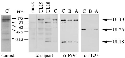
FIG. 2.
Analysis of capsid proteins. (Left panel) C capsids isolated from 60 to 20% sucrose gradients were subjected to SDS–12% PAGE and analyzed by Coomassie blue staining. (Right panels) Lysates of COS-7 cells transfected with pCG-UL19 (UL19) or pCG-UL18 (UL18) and gradient-purified C, B, and A capsids were analyzed by SDS–10% PAGE, followed by immunoblotting with anti-capsid, anti-PrV, or anti-UL25 antibodies as indicated below each blot. Positions of the UL19, UL18, and UL25 proteins are indicated on the right with respective arrows.

