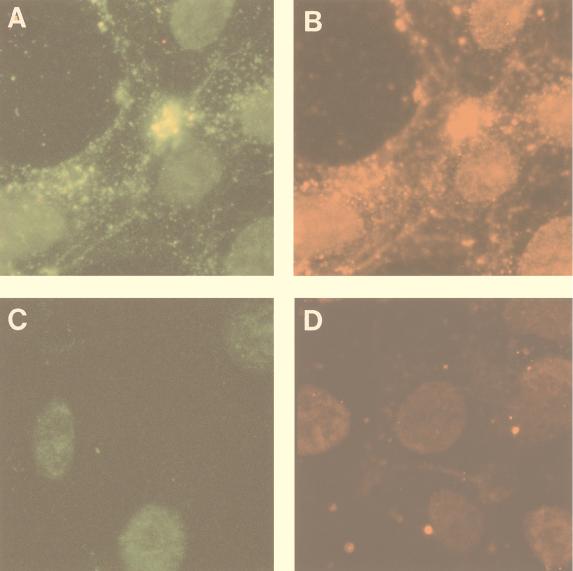
FIG. 6.
Immunofluorescence microscopy of incoming PrV nucleocapsids. COS-7 cells were either mock infected (C and D) or were infected with PrV at an MOI of 50 (A and B). Cells were extracted with 1% NP-40 at 30 min postinfection, fixed, and analyzed by double immunofluorescence with the rabbit anti-capsid antibody (A) and the mouse anti-UL25 antibody (B). To prevent occlusion of the UL25 sites on the capsid by the capsid sera, cells were incubated first with the UL25 antiserum alone and then with both antibodies. Mock-infected cells were analyzed with either the anti-capsid antibody (C) or anti-UL25 antibody alone (D). Secondary antibodies were donkey anti-rabbit antibody coupled to DTAF and anti-mouse antibody coupled to Texas red. Texas red and fluorescein signals were visualized by using narrow-band Texas red or fluorescein isothiocyanate filters. The same fields were photographed in panels A and B. Magnification, ×750.

