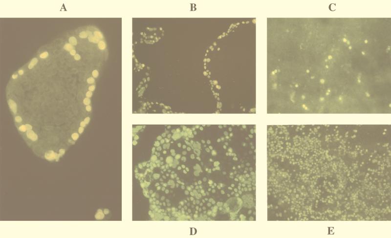
FIG. 1.
Cells were infected with CMV, fixed 48 h p.i., and examined by indirect immunofluorescence with antibody clone E13, directed against CMV IE proteins, and fluorescein isothiocyanate-conjugated secondary antibody. (A) Infection of 3-day-old Caco-2 cell islets appeared to be restricted to the outer edge. Six-day-old Caco-2 cells, which are subconfluent and consist of large islets, were infected with CMV without any treatment (B) or treated with EGTA (Sigma) (D). Fourteen-day-old Caco-2 cells grown on glass coverslips infected with CMV that were untreated (C) or treated with EGTA (E).

